
Dog Ultrasound
Dog Ultrasound
Outline of the Article:
- Introduction
- Understanding Dog Ultrasound
- Why Use Dog Ultrasound?
- The Benefits of Dog Ultrasound
- How Dog Ultrasound Works
- Preparing Your Dog for Ultrasound
- The Role of Veterinary Ultrasound Technicians
- Common Applications of Dog Ultrasound
- Advancements in Canine Ultrasound
- Interpreting Ultrasound Results
- Potential Risks and Limitations
- Cost of Dog Ultrasound
- Frequently Asked Questions (FAQs)
- Conclusion

Introduction
In the world of veterinary medicine, advancements in technology have brought about revolutionary diagnostic tools. One such tool is the use of ultrasound in dogs. Dog ultrasound, often used for diagnostic purposes, has become an essential part of veterinary care. In this article, we'll delve into the world of dog ultrasound, exploring its benefits, applications, and how it works. So, let's start our journey into the fascinating realm of canine ultrasound.
Understanding Dog Ultrasound
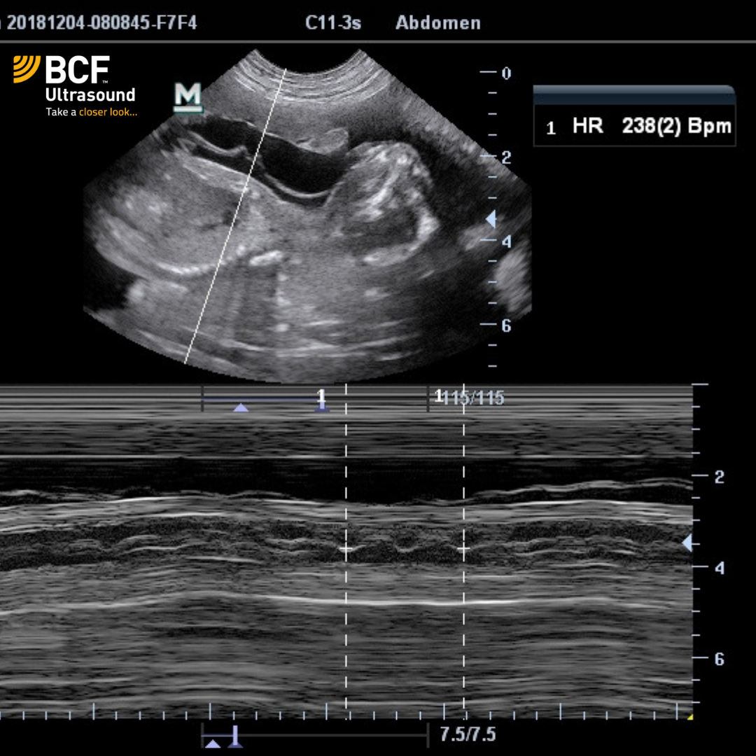
Dog ultrasound, also known as canine ultrasonography, is a non-invasive medical imaging technique that uses high-frequency sound waves to create real-time images of the internal structures of a dog's body. These images, called sonograms, are valuable for diagnosing and monitoring various health conditions in dogs.
To break it down further:
- Non-Invasive: Dog ultrasound is a non-invasive procedure, which means it doesn't require any surgical incisions or invasive methods to examine the internal organs and tissues of your dog. This is a significant benefit as it reduces the risk and discomfort associated with surgery.
- Real-Time Imaging: When a dog undergoes an ultrasound, veterinarians can immediately visualize the internal structures in real time. This allows for quick diagnosis and the ability to observe how the organs and tissues are functioning at that moment.
- Painless: The procedure is entirely painless for dogs. Unlike some other diagnostic methods, such as certain blood tests or endoscopy, ultrasound doesn't cause any pain or discomfort to your pet. This is especially important for keeping your dog at ease during the examination.
- Safe for Pregnancy: Ultrasound is commonly used for monitoring pregnant dogs and their developing puppies. It allows veterinarians to track the progress of the pregnancy, count the number of puppies, and ensure the health of both the mother and her offspring. This application has been a game-changer in breeding and prenatal care for dogs.
Dog ultrasound is a vital tool in veterinary medicine that enables veterinarians to peer inside a dog's body without the need for invasive procedures. It's non-invasive, painless, and provides real-time images, making it an invaluable resource for diagnosing and monitoring health conditions in our beloved canine companions.

Why Use Dog Ultrasound?
The Benefits of Dog Ultrasound
Dog ultrasound offers several advantages:
- Non-invasive: Unlike surgery, ultrasound doesn't require any incisions, making it a safer option for both diagnostic and monitoring purposes.
- Painless: The procedure is painless for dogs, ensuring their comfort during the examination.
- Real-time Imaging: Veterinarians can immediately see internal structures, enabling quick diagnosis and treatment.
- Safe for Pregnancy: Ultrasound is commonly used to monitor pregnant dogs and their developing puppies.
How Dog Ultrasound Works
Ultrasound machines emit high-frequency sound waves that bounce off internal structures in the dog's body. These waves are then recorded, creating detailed images. The density and type of tissue determine how the waves are reflected, resulting in varying shades on the ultrasound image.
Dog ultrasound, or canine ultrasonography, is a medical imaging technique that uses sound waves to create real-time images of the internal structures of a dog's body. Understanding how dog ultrasound works involves delving into the principles and technology behind this diagnostic method.
Here's a detailed explanation of how dog ultrasound works:
1. Sound Wave Generation:
The process begins with the ultrasound machine itself. This device contains a transducer, which is essentially a hand-held probe that emits high-frequency sound waves.
These sound waves are beyond the range of human hearing and are typically between 2 to 18 megahertz (MHz). The exact frequency used depends on the specific area being examined and the depth of the structures under investigation.
2. Sound Wave Transmission:
Once the transducer is placed on the dog's skin, it starts emitting these high-frequency sound waves.
The sound waves travel through the dog's skin and into the underlying tissues.
3. Sound Wave Interaction:
As the sound waves pass through the dog's body, they encounter different types of tissues, such as muscles, organs, and bones.
When the sound waves encounter a tissue boundary or a change in density, such as the boundary between muscle and a solid organ like the liver, part of the sound wave is reflected back towards the transducer while another part continues to penetrate deeper into the body.
4. Receiving and Interpretation:
The transducer also serves as a receiver, picking up the echoes or reflected sound waves.
These echoes are then sent to a computer in the ultrasound machine, which processes the information to create real-time images.
5. Image Formation:
The computer interprets the time it takes for the sound waves to bounce back and the intensity of the echoes to create images on the screen.
The images are grayscale, with different shades representing the density and composition of the tissues. For instance, fluids appear dark, while denser tissues like bone or organs appear as lighter shades.
6. Real-Time Imaging:
One of the significant advantages of dog ultrasound is that it provides real-time imaging. As the transducer is moved across the dog's body, the images on the screen change in response, allowing the veterinarian or technician to observe the structures and organs in motion.
7. Examination and Diagnosis:
The veterinarian examines these real-time images to diagnose various conditions, including abnormalities in organs, detecting pregnancies, or evaluating blood flow within the body.
By carefully studying the images, the veterinarian can make informed decisions about the dog's health.
8. No Harmful Radiation:
Unlike X-rays, which use ionizing radiation, ultrasound is non-ionizing and doesn't pose a risk of radiation exposure to the dog or the person performing the procedure.
Dog ultrasound works by emitting high-frequency sound waves into the dog's body, recording the echoes produced when these waves encounter different tissues and organs. The computer then processes these echoes into real-time images, providing invaluable insights for diagnosing and monitoring a dog's health without the need for invasive procedures or harmful radiation. This non-invasive, painless, and highly informative diagnostic method has become an indispensable tool in veterinary medicine.

Preparing Your Dog for Ultrasound
Before a dog ultrasound, it's essential to ensure your furry friend is well-prepared. This includes fasting for a specific period, ensuring the bladder is adequately filled, and maintaining a calm and cooperative demeanor.
Preparing your dog for an ultrasound is an essential step to ensure a successful and informative examination. Ultrasound is a non-invasive and painless procedure, but certain preparations are necessary to obtain clear images and make the experience as comfortable as possible for your furry friend.
Here's a detailed explanation of how to prepare your dog for an ultrasound:
1. Fasting:
In most cases, your veterinarian will ask you to fast your dog for a specific period before the ultrasound. Fasting typically involves withholding food for at least 8 to 12 hours before the procedure. Water, however, is usually not restricted, and your dog can continue to drink.
Fasting helps reduce the presence of gas and food in the digestive system, providing a clearer ultrasound image.
2. Bladder Preparation:
For abdominal ultrasounds, your veterinarian may instruct you to ensure that your dog's bladder is adequately filled before the examination. This is especially important for female dogs.
A full bladder helps in imaging pelvic and abdominal structures more effectively by pushing the organs into view and separating them from the surrounding tissue.
3. Comfort and Calmness:
It's crucial to ensure that your dog is comfortable and calm on the day of the ultrasound. Stress or anxiety can affect the dog's behavior and potentially the quality of the images.
Make sure your dog is relaxed and well-rested. You can bring along a favorite toy or blanket to provide comfort.
4. Cooperation:
During the ultrasound, your dog may need to be still for a certain period. If your dog is too anxious or active, it might be necessary to use mild sedation or anesthesia to ensure cooperation.
Discuss this with your veterinarian, and they will decide the best approach to keep your dog calm and still.
5. Hair Trimming:
Depending on the area to be examined, your veterinarian may need to trim or shave a small patch of your dog's fur. This allows for better contact between the ultrasound transducer and the skin, which is essential for obtaining clear images.
6. Consent and Questions:
Before the procedure, your veterinarian will typically discuss the purpose of the ultrasound, any potential risks, and the expected outcome. This is an opportunity to ask any questions you may have and provide consent for the examination.
7. Post-Procedure Care:
After the ultrasound, your dog may be a bit groggy if sedation or anesthesia was used. Provide a quiet and comfortable place for your dog to recover.
Follow any post-procedure care instructions given by your veterinarian, which may include resuming normal feeding and exercise routines.
Preparing your dog for an ultrasound involves fasting, ensuring a full bladder (if required), creating a calm and comfortable environment, and being prepared for the possibility of hair trimming or sedation. Proper preparation not only ensures the accuracy of the ultrasound but also contributes to a positive and stress-free experience for your canine companion. Your veterinarian will guide you through the specific preparations based on the purpose of the ultrasound and your dog's unique needs.
The Role of Veterinary Ultrasound Technicians
Veterinary ultrasound technicians play a crucial role in conducting the procedure. They are trained to operate the ultrasound machine, interpret the images, and assist veterinarians in diagnosing medical conditions.
Veterinary ultrasound technicians, often referred to as veterinary sonographers, play a crucial role in the field of veterinary medicine. They are highly skilled professionals responsible for operating ultrasound machines, conducting ultrasound examinations, and assisting veterinarians in diagnosing and monitoring medical conditions in animals, including dogs. Here, we'll explore the vital role that veterinary ultrasound technicians play in providing comprehensive care for animals.
1. Conducting Ultrasound Examinations:
One of the primary responsibilities of veterinary ultrasound technicians is to perform ultrasound examinations on animals. These examinations can be conducted for various purposes, including diagnosing illnesses, monitoring pregnancies, and evaluating organ health.
2. Operating Ultrasound Equipment:
Veterinary ultrasound technicians are experts in using ultrasound machines. They have a deep understanding of the technology, including how to adjust settings for different types of examinations and to obtain the best possible images.
3. Preparing Patients and Equipment:
Before an ultrasound examination, technicians ensure that both the patient (animal) and the equipment are well-prepared. This may involve shaving or trimming the fur in the examination area to facilitate better contact between the ultrasound transducer and the skin.
4. Assisting Veterinarians:
Veterinary ultrasound technicians work closely with veterinarians during examinations. They collaborate to obtain the necessary images and assist in making real-time assessments. Their expertise is invaluable for accurate and prompt diagnoses.
5. Image Interpretation:
While veterinary ultrasound technicians do not provide diagnoses themselves, they are trained to recognize anatomical structures and anomalies on ultrasound images. Their insights can help veterinarians focus on specific areas of concern.
6. Patient Comfort and Safety:
Ensuring the comfort and safety of the animal is a top priority for veterinary ultrasound technicians. They handle the animals gently, maintain a calming environment, and monitor the patient's well-being during the procedure.
7. Record-Keeping:
Technicians are responsible for documenting the ultrasound findings, including image descriptions and measurements. These records are crucial for patient histories and monitoring progress over time.
8. Continuing Education:
To stay current in their field, veterinary ultrasound technicians engage in ongoing education and training. They keep abreast of the latest advancements in ultrasound technology and diagnostic techniques.
9. Specialty Areas:
Some technicians may specialize in specific areas such as cardiac or abdominal ultrasound. This specialization allows them to offer a higher level of expertise in those particular areas.
10. Communication:
Effective communication with both the veterinary team and pet owners is essential. Veterinary ultrasound technicians often explain the procedure, findings, and any follow-up steps to the pet owner, ensuring they understand the diagnostic process and any necessary treatments.
Veterinary ultrasound technicians are highly skilled professionals who support veterinarians in the diagnosis and care of animals. Their role is multifaceted, encompassing the operation of ultrasound equipment, conducting examinations, assisting in image interpretation, and ensuring the well-being of the patients. Their expertise is indispensable in delivering comprehensive and effective healthcare to animals, making them an integral part of the veterinary team.

Common Applications of Dog Ultrasound
Dog ultrasound has a wide range of applications:
- Abdominal Imaging: It helps diagnose issues in the liver, kidneys, and gastrointestinal tract.
- Cardiac Ultrasound: Also known as echocardiography, it assesses the heart's structure and function.
- Pregnancy Monitoring: Ultrasound is used to track the development of fetuses during pregnancy.
Advancements in Canine Ultrasound
Advancements in technology have led to the development of more sophisticated ultrasound machines with improved image quality, better portability, and increased diagnostic accuracy. These advancements have made ultrasound an even more valuable tool in veterinary care.
Advancements in canine ultrasound have significantly enhanced the capabilities and diagnostic accuracy of this non-invasive imaging technique in veterinary medicine. These innovations have made ultrasound an indispensable tool for diagnosing and monitoring various medical conditions in dogs. Let's delve into the clear and exciting advancements in canine ultrasound.
1. Improved Image Quality:
One of the most notable advancements is the significant improvement in image quality. High-resolution imaging allows for clearer and more detailed visualization of internal structures, making it easier to detect abnormalities and provide accurate diagnoses.
2. Doppler Ultrasound:
Doppler ultrasound is an innovation that enables the assessment of blood flow within the dog's body. This is especially valuable in diagnosing cardiovascular conditions and monitoring blood flow in various organs.
3. 3D and 4D Imaging:
Three-dimensional (3D) and four-dimensional (4D) ultrasound have introduced a new dimension to canine imaging. These technologies provide veterinarians with a three-dimensional view of the dog's internal organs, offering a more comprehensive understanding of their structure and potential abnormalities.
4. Portable Ultrasound Machines:
Advancements have led to the development of portable ultrasound machines. These lightweight and compact devices are particularly beneficial for veterinarians working in the field, emergency situations, or in remote locations.
5. Color Flow Mapping:
Color flow mapping technology is used to visualize the direction and speed of blood flow. It's invaluable in diagnosing circulatory issues, such as blood clots or irregularities in the heart.
6. Tissue Harmonics Imaging:
Tissue harmonics imaging improves the quality of images in difficult-to-scan areas. It minimizes artifact interference, providing clearer images even in complex situations.
7. Elastography:
Elastography is a technique that assesses tissue stiffness. It aids in identifying abnormalities, particularly in soft tissues. This can be particularly helpful in detecting tumors and differentiating between benign and malignant masses.
8. Telemedicine and Image Sharing:
The integration of telemedicine allows for remote consultation and image sharing with specialists. Veterinarians can seek expert opinions and collaborate on challenging cases, leading to more accurate diagnoses and treatment plans.
9. Veterinary-Specific Software:
Specialized software packages are now available for veterinary ultrasound, tailored to the unique needs of diagnosing and treating dogs. These software tools assist in measurements, analysis, and documentation.
10. Training and Certification:
With these advancements, there has been a growing emphasis on the training and certification of veterinary ultrasound technicians, ensuring that they are proficient in using the latest technology and techniques.
11. Increased Accessibility:
The combination of portable devices and greater training opportunities has made ultrasound more accessible to a broader range of veterinary practices, including smaller clinics and rural areas.
Advancements in canine ultrasound have transformed the field of veterinary diagnostics. With improved image quality, innovative technologies, and increased accessibility, ultrasound has become an even more powerful tool for diagnosing and monitoring medical conditions in dogs. These advancements not only improve the accuracy of diagnoses but also contribute to the overall well-being of our canine companions by enabling prompt and effective medical care.

Interpreting Ultrasound Results
Interpreting ultrasound images requires expertise. Veterinarians assess the images and use them to make informed decisions about a dog's health. Clear and accurate interpretation is crucial for the correct diagnosis and treatment.
Interpreting ultrasound results is a crucial aspect of the diagnostic process in veterinary medicine. Ultrasound imaging provides detailed images of an animal's internal structures, allowing veterinarians to evaluate the health of various organs and tissues. Here, we'll explain how veterinarians interpret ultrasound results to make informed decisions about a dog's health.
1. Image Assessment:
The first step in interpreting ultrasound results is a thorough assessment of the images. Veterinarians carefully examine the ultrasound images to identify different organs, tissues, and structures within the dog's body.
2. Recognizing Normal Anatomy:
Veterinarians have a deep understanding of the normal anatomy of dogs. They compare the observed structures in the ultrasound images to what is expected in a healthy dog. This helps them identify any abnormalities or irregularities.
3. Measurements and Calculations:
In some cases, measurements of specific structures, such as the size of organs or the thickness of tissues, are taken. These measurements are compared to established reference ranges to assess whether they fall within normal limits.
4. Abnormalities and Pathology:
If abnormalities are detected, the veterinarian will seek to identify the nature of the issue. They consider factors such as the size, shape, and location of any masses or anomalies. For instance, a lump could be a benign cyst or a malignant tumor.
5. Blood Flow Assessment:
In cases where Doppler ultrasound is used, veterinarians assess blood flow patterns and velocity. Irregular blood flow can indicate issues such as blood clots, vascular problems, or heart conditions.
6. Integration with Clinical Data:
Ultrasound results are integrated with the dog's clinical history, physical examination findings, and other diagnostic tests (such as blood work or X-rays). This holistic approach helps veterinarians form a comprehensive understanding of the dog's health.
7. Differential Diagnosis:
The veterinarian considers various possible diagnoses based on the ultrasound findings and clinical information. They create a list of potential conditions and prioritize them based on the likelihood and severity of the disease.
8. Real-Time Assessment:
A significant advantage of ultrasound is the ability to perform real-time assessments. This means that veterinarians can observe the dog's internal structures in motion, allowing them to evaluate how organs function and respond to various stimuli.
9. Consultation and Collaboration:
In complex cases or when unusual findings are encountered, veterinarians may consult with specialists or colleagues for a second opinion. Collaboration and sharing of ultrasound images via telemedicine can aid in making more accurate diagnoses.
10. Communication with Pet Owners:
After interpreting the ultrasound results, the veterinarian communicates the findings and potential diagnoses to the pet owner. This discussion includes an explanation of the condition, its implications, and possible treatment options.
In conclusion, interpreting ultrasound results is a multi-faceted process that involves a thorough evaluation of images, a deep understanding of normal and abnormal anatomy, and the integration of clinical data. Veterinarians use their expertise to form diagnoses, prioritize potential conditions, and make informed decisions about the dog's healthcare. Effective communication with pet owners is also essential to ensure that they are well-informed and can actively participate in the treatment and care of their furry companions.
Potential Risks and Limitations
While dog ultrasound is generally safe, there are minimal risks associated with it. For instance, incorrect interpretation of images may lead to misdiagnosis. Additionally, some conditions may not be visible with ultrasound alone, requiring complementary diagnostic methods.
While canine ultrasound is a valuable and non-invasive diagnostic tool in veterinary medicine, it's essential to be aware of its potential risks and limitations. Understanding these factors can help veterinarians and pet owners make informed decisions when using ultrasound for the diagnosis and monitoring of dogs.
1. Operator Proficiency:
Ultrasound requires a skilled operator to obtain high-quality images and accurate results. Inexperienced or inadequately trained technicians may produce suboptimal images, which can lead to misdiagnosis or the need for additional imaging.
2. Limited Penetration:
Ultrasound has limitations in terms of the depth it can penetrate and the ability to visualize structures beyond a certain depth. This can be a constraint when assessing large or deep-seated organs.
3. Variability in Image Quality:
Image quality can vary based on factors like the dog's size, breed, age, and the presence of gas or other obstructions in the body. For example, gas in the gastrointestinal tract can limit ultrasound's effectiveness.
4. Patient Cooperation:
Ultrasound typically requires the dog to remain still during the examination. An anxious or uncooperative dog may require sedation or anesthesia, which can introduce risks and additional costs.
5. Complementary Diagnostics:
While ultrasound is a powerful tool, it may not provide a complete picture on its own. Veterinarians may need to combine ultrasound findings with other diagnostic tests like X-rays, blood work, or biopsies to arrive at a definitive diagnosis.
6. Interpretation Challenges:
Interpreting ultrasound images can be complex, especially in cases with subtle or unusual findings. Differentiating between benign and malignant masses, for example, may be challenging and require further evaluation.
7. Operator-Dependent:
The accuracy of ultrasound results depends on the operator's skill and experience. Variability in proficiency among different technicians can impact the reliability of the examination.
8. Overdiagnosis or Underdiagnosis:
There's a potential for both overdiagnosis and underdiagnosis. Overly cautious interpretations may lead to unnecessary treatments, while underestimating the severity of a condition may delay essential interventions.
9. Limited Use for Bone and Air Evaluation:
Ultrasound is less effective for evaluating bones and air-filled structures. To assess these areas, other imaging modalities like X-rays or CT scans are necessary.
10. Inability to Monitor Some Organs:
While ultrasound is excellent for visualizing soft tissues and organs like the liver, kidneys, and heart, it may not provide sufficient information for monitoring certain organs, such as the brain.
11. False Negatives:
In some cases, ultrasound may yield false-negative results, failing to detect abnormalities that are present but not visible due to various factors.
12. Additional Costs:
Ultrasound, while a valuable tool, can add to the overall cost of veterinary care. In cases where specialized or repeated ultrasounds are needed, these costs can be substantial.
13. Risk of Stress or Discomfort:
The ultrasound examination process may cause stress or discomfort to some dogs, particularly if they are anxious or if the examination area is sensitive. Efforts to minimize these risks are essential for the well-being of the animal.
While canine ultrasound is a highly valuable and safe diagnostic tool, it is not without its limitations and potential risks. Veterinarians must consider these factors when recommending and interpreting ultrasound examinations. Collaborative decision-making between veterinarians and pet owners is essential to ensure the appropriate and effective use of ultrasound in diagnosing and caring for dogs.
Cost of Dog Ultrasound
The cost of dog ultrasound varies depending on factors like the purpose of the procedure, the location, and the complexity of the case. It's essential to discuss pricing with your veterinarian before proceeding.
The cost of a dog ultrasound can vary based on several factors, including the purpose of the procedure, the location of the veterinary clinic, and the complexity of the case. Understanding the cost of dog ultrasound is important for pet owners to budget for veterinary care effectively. Here's a detailed explanation of the factors that influence the cost of a dog ultrasound:
1. Purpose of the Ultrasound:
The purpose of the ultrasound significantly impacts the cost. Routine wellness ultrasounds, which are performed as part of a regular check-up, are generally less expensive than diagnostic ultrasounds conducted to investigate specific health concerns. Pregnancy monitoring ultrasounds may also have a different pricing structure.
2. Geographic Location:
The cost of veterinary services, including ultrasounds, can vary by location. In urban areas, where the cost of living is higher, veterinary services tend to be more expensive than in rural or less populated areas.
3. Type of Ultrasound:
Different types of ultrasounds may be required for various purposes. For example, abdominal ultrasounds are commonly used to assess organs in the abdomen, while cardiac ultrasounds focus on the heart. The type of ultrasound can affect the cost.
4. Complexity and Time Required:
The complexity of the case and the time required for the procedure can influence the cost. For instance, if the ultrasound involves a comprehensive examination of multiple organs or the need for multiple scans, it may be more expensive.
5. Need for Sedation or Anesthesia:
Some dogs may require sedation or anesthesia during the ultrasound to ensure they remain still and cooperative. The use of sedatives or anesthesia adds to the overall cost of the procedure.
6. Specialist Involvement:
In cases where a veterinary specialist, such as a veterinary radiologist or cardiologist, is involved in the interpretation of the ultrasound, there may be an additional fee for their expertise.
7. Follow-Up Tests:
Depending on the findings of the ultrasound, additional tests or procedures may be required, such as biopsies, blood work, or further imaging. These additional tests come with their own costs.
8. Veterinary Clinic and Reputation:
The reputation and experience of the veterinary clinic can also affect the cost. Established and well-regarded clinics may charge more for their services.
9. Pet Insurance:
Pet insurance can help offset the cost of veterinary procedures, including ultrasounds. Depending on the insurance policy, it may cover a portion of the ultrasound expenses.
10. Discounts or Packages:
Some veterinary clinics offer discounted packages for routine ultrasounds or comprehensive wellness examinations, which can provide cost savings for pet owners.
11. Emergency or After-Hours Services:
If the ultrasound is required outside of regular clinic hours, such as in emergencies, it may incur additional fees.
The cost of a dog ultrasound is influenced by various factors, including the purpose of the ultrasound, geographic location, type of ultrasound, complexity of the case, the need for sedation or anesthesia, specialist involvement, and follow-up tests. It's essential for pet owners to discuss the expected costs with their veterinarian and inquire about potential discounts or package deals to manage expenses effectively while ensuring the best care for their furry companions.
Frequently Asked Questions (FAQs)
1. Is dog ultrasound painful for my pet?
No, dog ultrasound is a painless and non-invasive procedure, and your pet will not experience any discomfort during the examination.
2. How long does a typical dog ultrasound take?
The duration of a dog ultrasound can vary, but it usually takes 20 to 45 minutes, depending on the purpose and complexity of the examination.
3. Can dog ultrasound detect all medical conditions?
While dog ultrasound is a powerful diagnostic tool, it may not detect every medical condition. Some issues may require other diagnostic methods like X-rays or blood tests.
4. Is anesthesia required for a dog ultrasound?
In most cases, anesthesia is not necessary for a dog ultrasound. However, it may be needed for specific situations or for very anxious dogs.
5. How often should I have my dog undergo ultrasound?
The frequency of dog ultrasounds depends on your dog's health and any specific concerns. Your veterinarian will recommend the appropriate schedule.
Conclusion
Dog ultrasound is a remarkable medical tool that has transformed the world of veterinary care. Its non-invasive nature, real-time imaging, and ability to diagnose a wide range of conditions make it invaluable for the health and well-being of our canine companions. As a responsible pet owner, it's essential to consider ultrasound when seeking the best possible care for your dog's health.
Dog ultrasound is not only a diagnostic method but also a gesture of love and care for your furry friend. By understanding the benefits and applications of this technology, you can ensure that your dog receives the best possible healthcare.
Only a few particularly fearful dogs may require a sedative. This is the reference examination.
 Small Animal Abdominal Ultrasonography Part 1 A Tour Of The Abdomen Today S Veterinary Practice
An ultrasound also called a sonogram is a non invasive procedure used to evaluate the internal organs in dogs and other animals
- Sound Wave Generation: The procedure begins with the ultrasound machine, which houses a hand-held device called a transducer. This transducer emits high-frequency sound waves, often beyond the range of human hearing, into the dog's body.
- Sound Wave Interaction: As these sound waves travel through the dog's body, they encounter different tissues and organs. When they come across a boundary between tissues or a change in density, such as the transition between muscle and an organ, some of the sound waves are reflected back toward the transducer, while others continue to penetrate deeper.
- Receiving and Interpretation: The transducer not only emits sound waves but also functions as a receiver. It picks up the echoes or reflected sound waves. These echoes are then sent to a computer within the ultrasound machine, which processes the information to create real-time images on a screen.
- Image Formation: The computer interprets the time it takes for the sound waves to bounce back and the intensity of the echoes. These factors generate detailed grayscale images that display the density and composition of the tissues and structures. Fluids appear dark, whereas denser tissues like bones or organs appear as lighter shades.
- Real-Time Imaging: What sets ultrasound apart is its ability to provide real-time imaging. As the transducer is moved across the dog's body, the images on the screen change dynamically, enabling veterinarians to observe the internal structures in motion. This is particularly valuable for assessing organ function and observing abnormalities in real time.
- Diagnostic Imaging: Ultrasound is an essential tool for diagnosing and monitoring various medical conditions, such as detecting tumors, evaluating organ health, and identifying abnormalities in the abdomen, heart, and other areas.
- Pregnancy Monitoring: In the field of breeding, ultrasound plays a crucial role in monitoring pregnancies. It allows veterinarians to confirm the presence of embryos or fetuses, count the number of offspring, and assess their viability.
- Cardiac Assessment: Cardiac or heart ultrasounds, also known as echocardiograms, are used to examine the heart's structure and function, aiding in the diagnosis of cardiac diseases and conditions.
- Guidance for Procedures: Ultrasound is used to guide procedures such as fine-needle aspirates or biopsies, providing real-time visualization to ensure the correct location for sample collection.
- Monitoring and Follow-Up: Ultrasound is valuable for tracking the progression of diseases, monitoring the response to treatments, and assessing the healing process after surgeries.
Dog Ultrasound Prenatal Care Ultrasound Pictures Ultrasound
 Using Ultrasound To Evaluate The Canine Pregnancy


